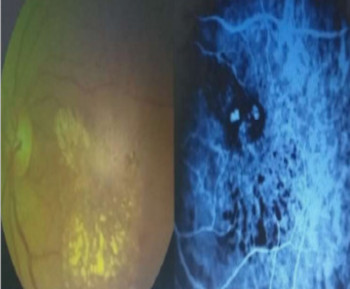
Panretinal photocoagulation: Indications, outcomes, and complications in retinal ischemic disorders at a tertiary eye care center in Kano State
Keywords:
Panretinal photocoagulation, proliferative diabetic retinopathy, retinal ischemia , laser photocoagulation, visual outcomeAbstract
INTRODUCTION
Panretinal photocoagulation (PRP) remains the gold standard for managing proliferative diabetic retinopathy (PRP) and other retinal ischemic disorders.1,2 By applying laser burns to the peripheral retina, PRP reduces oxygen demand and promotes regression of neovascularization.3,4 While its efficacy in reducing the risk of severe vision loss is well established, PRP may be associated with adverse effects that impact visual outcomes.5,6,7 The objective of this study was to describe the indications, visual outcomes, and complications associated with PRP in patients treated at a tertiary eye care center.
METHODS
A retrospective review was conducted of patients who underwent PRP at Makkah Eye Specialist Hospital, Kano over a period of two years (July 2022 to June 2024). Data extracted included demographic characteristics, clinical indications, number of PRP sessions, visual acuity (pre-and post-treatment), complications, and need for adjunctive therapy. Descriptive analysis was performed using IBM SPSS version 26.
RESULTS
A total of 61 eyes of 33 patients (mean age: 51.1 ± 10.3 years, 69.7% male) were included. The most common indication was Proliferative diabetic retinopathy (39.4%), followed by Advanced diabetic eye disease in 27.3% of patients (Table 1). Visual acuity improved in 23.0% of patients, remained stable in 57.4%, and worsened in 19.6% (Table 2). Reported complications included Vitreous hemorrhage (8.3 %), and Tractional retinal detachment (5.0 %). Anti-VEGF therapy was required in 5.0% of cases, while 25.8% of patients underwent subsequent vitrectomy.
CONCLUSION
PRP is effective in stabilizing vision and slowing disease progression in ischemic retinopathies. Majority of patients achieve visual stability, although some may require adjunctive interventions. Close monitoring is essential to manage potential complications and optimize outcomes.
References
1. Diabetic Retinopathy Study Research Group; Indications for photocoagulation treatment of diabetic retinopathy. Diabetic Retinopathy Study report No; 14 Int Ophthalmol Clin 1987;27:239-53
2. The Central Retinal Vein Occlusion Group. A randomized clinical trial of early panretinal photocoagulation for ischemic central vein occlusion. The central vein occlusion study group N report. Ophthalmology 1995; 102:1432-44.
3. David B. Karlin (ed). Lasers in Opthalmic Surgery. Blackwell Science Inc.1995.
4. Steven M. Bloom, Alexander J Brucker (eds). Laser Surgery of the Posterior Segment (2nd ed). Philadelphia: Lippincott-Raven 1997.
5. Fu DJ, Thottarath S, Faes L, et al. Visual acuity outcome of stable proliferative diabetic retinopathy following initial complete panretinal photocoagulation. BMJ Open Ophthalmology. 2022; 7:e001068 PMC.
6. Yoshihiro Y, et al. American Society of Retina Specialists Clinical Practice Guidelines; Management of Nonproliferative and Proliferative Diabetic Retinopathy Without Diabetic Macular Edema. J Vitreoretin Dis. 2020 Jan 6;4(2):125-135.
7. Antoszyk AN, Glassman AR, Beaulieu WT et al. Effect of Intravitreous Aflibercept vs Vitrectomy With Panretinal Photocoagulation on Visual Acuity in Patients With Vitreous Hemorrhage From Proliferative Diabetic Retinopathy: A Randomized Clinical Trial. JAMA 2020 ;324(23):2383-2395.
Additional Files
Published
How to Cite
Issue
Section
Categories
License
Copyright (c) 2025 Transactions of the Ophthalmological Society of Nigeria

This work is licensed under a Creative Commons Attribution-NonCommercial-NoDerivatives 4.0 International License.